Computed tomography (CT) of the body uses special x-ray equipment to help detect a variety of diseases and conditions. CT scanning is fast, painless, noninvasive and accurate. In emergency cases, it can reveal internal injuries and bleeding quickly enough to help save lives.
In many ways CT scanning works very much like other x-ray examinations. Different body parts absorb the x-rays in varying degrees. It is this crucial difference in absorption that allows the body parts to be distinguished from one another on an x-ray film or CT electronic image.
In a conventional x-ray exam, a small amount of radiation is aimed at and passes through the part of the body being examined, recording an image on a special electronic image recording plate. Bones appear white on the x-ray; soft tissue, such as organs like the heart or liver, shows up in shades of gray, and air appears black.
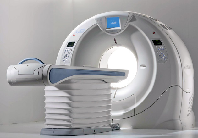
With CT scanning, numerous x-ray beams and a set of electronic x-ray detectors rotate around you, measuring the amount of radiation being absorbed throughout your body. Sometimes, the examination table will move during the scan, so that the x-ray beam follows a spiral path. A special computer program processes this large volume of data to create two-dimensional cross-sectional images of your body, which are then displayed on a monitor. CT imaging is sometimes compared to looking into a loaf of bread by cutting the loaf into thin slices. When the image slices are reassembled by computer software, the result is a very detailed multidimensional view of the body's interior.
Refinements in detector technology allow nearly all CT scanners to obtain multiple slices in a single rotation. These scanners, called multislice CT or multidetector CT, allow thinner slices to be obtained in a shorter period of time, resulting in more detail and additional view capabilities.
CT imaging is:
one of the fastest and most accurate tools for examining the chest, abdomen and pelvis because it provides detailed, cross-sectional views of all types of tissue.used to examine patients with injuries from trauma such as a motor vehicle accident.performed on patients with acute symptoms such as chest or abdominal pain or difficulty breathing.often the best method for detecting many different cancers, such as lymphoma and cancers of the lung, liver, kidney, ovary and pancreas since the image allows a physician to confirm the presence of a tumor, measure its size, identify its precise location and determine the extent of its involvement with other nearby tissue.an examination that plays a significant role in the detection, diagnosis and treatment of vascular diseases that can lead to stroke, kidney failure or even death. CT is commonly used to assess for pulmonary embolism (a blood clot in the lung vessels) as well as for aortic aneurysms.invaluable in diagnosing and treating spinal problems and injuries to the hands, feet and other skeletal structures because it can clearly show even very small bones as well as surrounding tissues such as muscle and blood vessels.
In pediatric patients, CT imaging is often used to evaluate:
lymphomaneuroblastomakidney tumorscongenital malformations of the heart, kidneys and blood vesselscystic fibrosiscomplications of acute appendicitiscomplications of pneumoniainflammatory bowel diseasesevere injuries
Radiologists and radiation oncologists often use the CT examination to:
quickly identify injuries to the lungs, heart and vessels, liver, spleen, kidneys, bowel or other internal organs in cases of trauma.guide biopsies and other procedures such as abscess drainages and minimally invasive tumor treatments.plan for and assess the results of surgery, such as organ transplants or gastric bypass.stage, plan and properly administer radiation treatments for tumors as well as monitor response to chemotherapy.measure bone mineral density for the detection of osteoporosis.
You should wear comfortable, loose-fitting clothing to your exam. You may be given a gown to wear during the procedure.
Metal objects, including jewelry, eyeglasses, dentures and hairpins, may affect the CT images and should be left at home or removed prior to your exam. You may also be asked to remove hearing aids and removable dental work. Women will be asked to remove bras containing metal underwire. You may be asked to remove any piercings, if possible.
You will be asked not to eat or drink anything for a few hours beforehand, as contrast material will be used in your exam. You should inform your physician of all medications you are taking and if you have any allergies. If you have a known allergy to contrast material, or "dye," your doctor may prescribe medications (usually a steroid) to reduce the risk of an allergic reaction. These medications generally need to be taken 12 hours prior to administration of contrast material. To avoid unnecessary delays, contact your doctor before the exact time of your exam.
Also inform your doctor of any recent illnesses or other medical conditions and whether you have a history of heart disease, asthma, diabetes, kidney disease or thyroid problems. Any of these conditions may increase the risk of an unusual adverse effect.
Women should always inform their physician and the CT technologist if there is any possibility that they may be pregnant.
Lorem Ipsum is simply dummy printing and typesetting industry. when an unknown printer took a galley of type and scrambled.